
Best overall: clinician-interpreted digital trichoscopy. Best for tracking growth: a phototrichogram. Best for suspected scarring: a scalp biopsy. The best hair loss diagnostic test in 2026 depends on the question your doctor needs to answer—not how advanced the scanner sounds.
- The best hair loss diagnostic test for visible scalp patterns is clinician-interpreted digital trichoscopy.
- Phototrichograms measure changes over time; targeted blood tests investigate specific suspected contributors.
- Scalp biopsy helps investigate suspected scarring or an uncertain diagnosis, not routine shedding.
- iGraft offers computerized hair assessments; ask how findings connect to your diagnosis and treatment plan.
Why this matters
A scalp scan, blood test and biopsy answer different questions. Buying a package of tests without a clinical examination risks giving you more results without explaining your hair loss.
iGraft offers computerized hair checkups, follicular-level scans and doctor-led assessments. iGraft hair loss diagnostic assessments are best suited to people seeking an in-person examination alongside computerized scalp findings. Ask which examination method is being used and what the report can establish; a computerized report is not, by itself, a diagnosis.
For your 2026 assessment, the useful outcome is a working diagnosis, an explanation of uncertainty and a plan for what happens next. Treatment selection comes after that—not before.
What makes the best hair loss diagnostic test?
Judge a diagnostic method against these criteria before comparing equipment or booking a package:
- Clinical fit: Does the test investigate your actual symptoms—shedding, patterned thinning, patches or scalp inflammation?
- Diagnostic contribution: Does it help distinguish plausible causes, rather than simply document less hair?
- Repeatability: Can the same area be assessed consistently at follow-up?
- Interpretation: Will a qualified clinician connect the result with your history and examination?
- Burden: Does the information justify clipping hair, taking blood or removing a tissue sample?
- Actionability: Will the result change treatment, monitoring or referral?
No single method wins every category. A detailed imaging report still cannot answer a question that requires blood testing or tissue examination.
Hair loss diagnostic tests at a glance
| Rank and method | Best for | Standout contribution | Key limitation |
|---|---|---|---|
| 1. Digital trichoscopy | Assessing visible hair and scalp patterns | Magnified examination of shafts, follicular openings and scalp features | Findings need clinical interpretation |
| 2. Phototrichogram | Measuring change over time | Repeat imaging of a defined scalp area | Requires careful positioning and preparation |
| 3. Hair pull test | Checking active shedding during examination | Immediate bedside assessment without imaging equipment | Does not identify the cause alone |
| 4. Targeted blood tests | Investigating suspected systemic contributors | Tests a specific clinical suspicion | Normal results do not exclude scalp disease |
| 5. Scalp biopsy | Investigating suspected scarring or unresolved diagnoses | Microscopic examination of scalp tissue | Invasive and dependent on sample selection |
Only the first two options are computerized imaging methods. The others belong in this comparison because a computer cannot replace the diagnostic information they provide.
1. Digital trichoscopy: best for an initial scalp-pattern assessment
Digital trichoscopy uses magnified scalp images to examine hair shafts, follicular openings and surrounding skin. A clinician assesses features such as differences in hair thickness, broken hairs, scaling and signs of inflammation alongside your symptoms.
Best for: People with patterned thinning, patchy loss or scalp changes that need closer examination.
Digital trichoscopy earns the default recommendation because it adds visible detail without removing tissue. DermNet’s clinical reference on trichoscopy describes its use across several hair and scalp disorders; its value lies in interpreting patterns, not generating a universal hair-health score.
Digital trichoscopy pros:
- Examines scalp detail that ordinary photographs miss.
- Provides images that can support explanation and follow-up.
- Helps clinicians select areas requiring closer investigation.
Digital trichoscopy cons:
- Overlapping findings do not always establish a diagnosis.
- Images cannot establish blood nutrient levels or thyroid function.
A computerized scan is not automatically a documented trichoscopy examination. For an iGraft hair loss diagnostic assessment, ask whether the report includes magnified images, which findings the doctor identifies and whether further testing is necessary.
Verdict: Skip image-only reports that provide a treatment recommendation without clinical interpretation.
2. Phototrichogram: best for measuring treatment-related change
A phototrichogram records a defined scalp area and uses repeat images to assess hair growth characteristics. Some protocols clip a small patch and photograph it again after a specified interval, commonly 48–72 hours, to distinguish growing from non-growing hairs.
Best for: People who need structured monitoring rather than another impression of whether their hair looks thicker.
Computer-assisted analysis can quantify features such as hair density and shaft diameter within the sampled area. The result describes that area; it does not automatically represent your entire scalp or explain why hair loss started.
Phototrichogram pros:
- Produces measurements suitable for repeat comparison.
- Separates monitoring from memory or casual photographs.
- Helps document change in a selected area.
Phototrichogram cons:
- Clipping and repeat appointments add practical demands.
- Different locations, preparation or software settings weaken comparisons.
For a 2026 monitoring plan, ask how the clinic will relocate the same site and keep imaging conditions consistent. A graph is useful only when its measurements are comparable.
Verdict: Hold claims of treatment success until follow-up measurements use a consistent protocol.
3. Hair pull test: best for checking active shedding
A hair pull test assesses whether hairs release readily when a clinician applies gentle traction to a small bundle. The finding helps assess active shedding, but interpretation depends on the examination technique and recent hair-care history.
Best for: People reporting increased shedding who need a bedside assessment alongside their history.
This is not a computerized test, and that is its advantage: it answers a focused question without requiring a scanner. A positive result does not identify the cause, and a negative result does not rule out every hair-loss disorder.
Hair pull test pros:
- Adds information during the clinical examination.
- Requires no imaging equipment or tissue removal.
- Helps connect reported shedding with an observable finding.
Hair pull test cons:
- Technique and recent washing affect interpretation.
- Cannot independently distinguish all causes of shedding.
Bring a clear timeline of illness, medication changes, childbirth, weight changes and new scalp symptoms. If your concern is female-pattern thinning rather than shedding alone, the guide to hair loss treatments for women helps frame the treatment discussion after diagnosis.
Verdict: Skip repeated self-testing; let a clinician interpret shedding in context.
4. Targeted blood tests: best for suspected internal contributors
Blood tests investigate specific clinical suspicions, such as iron deficiency or thyroid dysfunction. Depending on your history and examination, a doctor can select tests such as a full blood count, ferritin or thyroid function testing.
Best for: People whose symptoms, medical history or shedding pattern suggest an internal contributor.
A blood panel does not photograph follicles or distinguish every scalp disorder. Equally, a scalp scan cannot confirm a deficiency; these methods answer different questions and sometimes belong in the same assessment.
Targeted blood test pros:
- Investigates suspected contributors beyond the scalp.
- Provides results that can guide further medical assessment.
- Supports treatment of an identified abnormality when clinically appropriate.
Targeted blood test cons:
- Broad screening can produce findings unrelated to the hair complaint.
- A normal panel does not exclude patterned or inflammatory hair loss.
Ask what each requested test is investigating and how its result would change the plan. Do not start supplements solely because hair is shedding; the diagnosis and your wider health determine whether supplementation is appropriate.
Verdict: Skip blanket deficiency packages without an explained clinical reason.
5. Scalp biopsy: best for suspected scarring hair loss
A scalp biopsy removes a small tissue sample for microscopic examination. It is useful when scarring hair loss is suspected or the diagnosis remains uncertain after clinical examination and imaging.
Best for: People whose dermatologist needs tissue-level information to distinguish competing diagnoses.
A commonly used scalp punch biopsy is 4 mm in diameter, although the clinician selects the sampling approach. DermNet’s scalp biopsy guidance explains why the sampling site and tissue processing matter; the presence of a biopsy report does not remove those practical considerations.
Scalp biopsy pros:
- Examines structures below the visible scalp surface.
- Helps distinguish inflammatory and scarring processes.
- Can resolve diagnostic uncertainty that imaging leaves unanswered.
Scalp biopsy cons:
- Requires local anaesthetic and wound care and leaves a small scar.
- An unsuitable sampling site can limit diagnostic information.
Do not delay assessment of progressive patches accompanied by pain, burning, redness or apparent loss of follicular openings. Those findings need clinical attention rather than another consumer scan.
Verdict: Hold cosmetic procedure decisions until suspected scarring or inflammation has been assessed.
How to get a useful diagnostic appointment
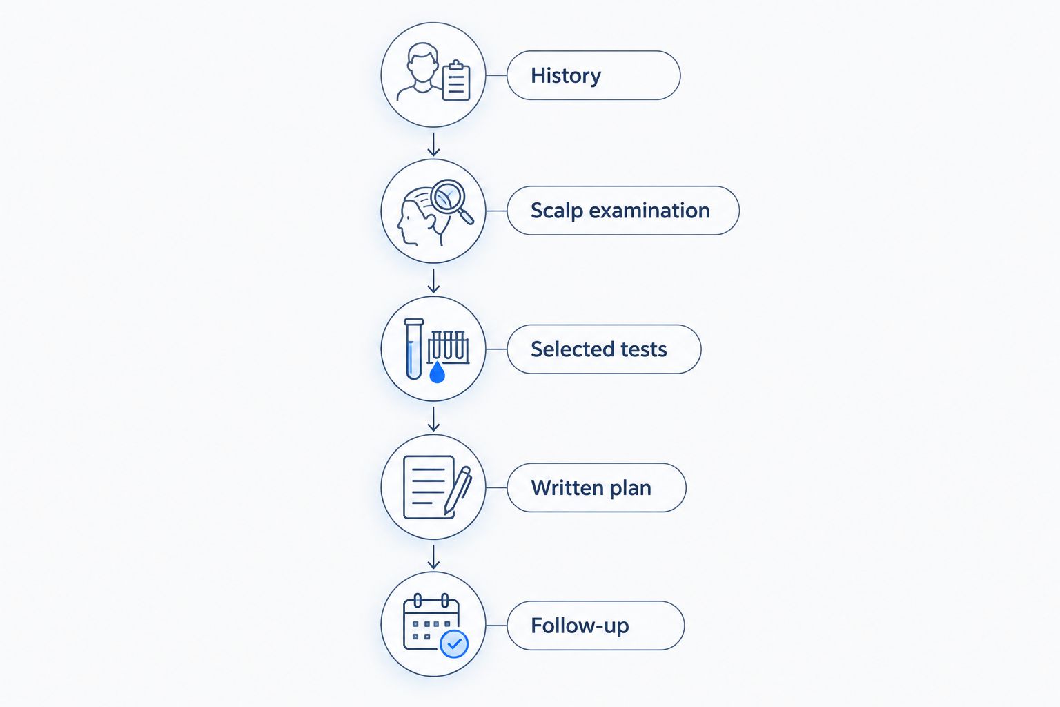
A productive appointment follows a clinical sequence, not a technology sales pitch. Use these steps to keep the discussion focused:
- History: Describe onset, progression, shedding, symptoms, medicines and relevant health changes.
- Scalp examination: Ask which findings support the proposed diagnosis and which alternatives remain possible.
- Selected tests: Choose imaging, blood tests or biopsy only where they answer an unresolved question.
- Written plan: Record the diagnosis, next action and what would trigger reassessment.
- Follow-up: Agree how progress will be measured under comparable conditions.
This sequence also separates diagnosis from treatment promotion. An impressive scan should not move you straight into a procedure discussion before the clinician explains what is happening.
For your 2026 consultation, bring previous reports and a dated symptom timeline rather than relying on memory. Ask these 3 questions before agreeing to treatment: What is the working diagnosis? What evidence supports it? What would change the plan?
Start with a clinical assessment
Discuss your hair-loss history, scalp findings and the next diagnostic step.
How these tests are ranked
This ranking prioritizes clinical fit, diagnostic contribution, repeatability, interpretation, burden and actionability. It is a guide to choosing the right diagnostic question—not a claim that every person needs every test.
Digital trichoscopy leads for a closer initial look at visible patterns. Phototrichograms lead for structured monitoring, while targeted blood tests and biopsy move ahead when the clinical question specifically requires them.
Which hair loss diagnostic test should you choose?
Start with a clinician-led history and scalp examination; add digital trichoscopy when magnification helps explain the findings. That is the practical default for someone comparing computerized hair assessments in 2026.
Choose a phototrichogram when the priority is repeat measurement. Choose targeted blood testing for a specific suspected contributor, and follow a dermatologist’s advice about biopsy when scarring or unresolved inflammation is a concern.
At iGraft, ask for an explanation connecting the computerized assessment to the proposed diagnosis. The report should help you understand the decision, not substitute for the consultation.
FAQ
What’s the best hair loss diagnostic test in 2026?
Clinician-interpreted digital trichoscopy is a useful default for examining visible hair and scalp patterns. The best test changes when the question concerns systemic contributors, measured growth or suspected scarring.
Is a computerized hair scan enough to diagnose hair loss?
No, a computerized hair scan is not enough on its own. A clinician must interpret the images alongside your history and examination, then decide whether additional testing is needed.
Is a phototrichogram better than trichoscopy?
A phototrichogram is better suited to repeat measurement, while trichoscopy examines diagnostic patterns in hair and scalp features. Neither replaces the other in every situation.
Do I need blood tests for hair fall?
You need blood tests when your history or examination gives a clinical reason to investigate an internal contributor. A doctor should explain which suspected condition each test addresses.
When is a scalp biopsy necessary for hair loss?
A scalp biopsy is useful when a dermatologist suspects scarring hair loss or needs tissue examination to clarify an uncertain diagnosis. It is not a routine requirement for every person with shedding.
What should I ask about an automated hair report?
Ask which findings a clinician has confirmed and how they support the diagnosis. Also ask what the software cannot determine and whether follow-up images will use the same method.
Can a diagnostic scan tell me which treatment will work?
A diagnostic scan cannot guarantee a treatment response. Imaging contributes to diagnosis and monitoring, while treatment selection also depends on the condition, medical history and clinical assessment.
One last thing
A more detailed report is not automatically a more useful report. Before leaving your 2026 assessment, ask the clinician to explain what would change the working diagnosis; that answer shows whether the plan accounts for uncertainty rather than hiding it behind a score.
Related guides